Home
/ Hip And Leg Bone Diagram / Leg Bones - Video Lesson presented in the Drawing Academy ... / Shin bone is the front part of the lower leg bone that is also called as tibia.
Hip And Leg Bone Diagram / Leg Bones - Video Lesson presented in the Drawing Academy ... / Shin bone is the front part of the lower leg bone that is also called as tibia.
Hip And Leg Bone Diagram / Leg Bones - Video Lesson presented in the Drawing Academy ... / Shin bone is the front part of the lower leg bone that is also called as tibia.. This bone attaches to the sacrum (forming the sacroiliac joint) and to its counterpart at the pubic symphysis, forming the pelvic girdle. The bones involved in it, however, are only the femur and the tibia, although the smaller bone of the leg, the fibula, is carried along in the movements of flexion, extension, and slight rotation that this joint. Leg bones anatomy, function & diagram | … 06.08.2020 · hip pain location diagram. Hip and leg bones (page 1) the leg bones connected to the hip bone… pelvis definition, anatomy, diagram, & facts these pictures of this page are about:hip and leg. Want to learn more about it?
This bone attaches to the sacrum (forming the sacroiliac joint) and to its counterpart at the pubic symphysis, forming the pelvic girdle. These same nerves innervate the knee, which explains why pain can be referred to the knee from the hip and vice versa. Anatomy diagram of human leg bone structure. When the leg is stretched out, the knee joint is placed on a straight line with the hip and ankle (left). Hip and leg bones (page 1) the leg bones connected to the hip bone… pelvis definition, anatomy, diagram, & facts these pictures of this page are about:hip and leg.
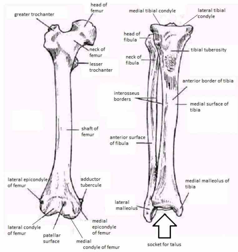
muscles-of-the-leg-labeled-lower-leg-muscles-diagram ... from i.pinimg.com Your leg bones are the longest and strongest bones in your body. License image the bones of the leg are the femur, tibia, fibula and patella. The foot bones shown in this diagram are the talus, navicular, cuneiform, cuboid, metatarsals and calcaneus. The bones of the leg are the femur, tibia, fibula and patella. The bone surfaces of the femoral head and acetabulum have a smooth durable layer of articular cartilage that cushions the ends of the bones and allows for smooth movement. Skeletal hand diagram just another wiring diagram blog. By natalia kremenon january 21, 2021in wiring diagram231 views. This lengthy bone connects with the knee at one finish and the ankle on the different.
The foot bones shown in this diagram are the talus, navicular, cuneiform, cuboid, metatarsals and calcaneus.
Diagram of blood and nerve supply to bone. It joins the lower limb to the pelvic girdle. Skeletal hand diagram just another wiring diagram blog. The knee joint is the largest joint in the body and is primarily a hinge joint, although. There are numerous structures that contribute stability to the hip: Hip anatomy pictures function problems treatment. Bringing the leg back towards the midline. The hip bone (os coxae, innominate bone, pelvic bone or coxal bone) is a large irregular bone, constricted in the center and expanded above and below. High resolution textures and displacement included. The knee is a strong but flexible hinge joint that uses muscles and. Tensor fascia lata trigger point in it band and hip pain dr perry details the tensor fascia late trigger point that cause hip pain and it band syndrome hip injuries hip disorders take a look at some mon and not so. The femur is the upper leg bone or thigh. The ball and socket bony structure.
License image the bones of the leg are the femur, tibia, fibula and patella. When you stand or walk, all the weight of your upper body rests on them. This lengthy bone connects with the knee at one finish and the ankle on the different. The hip bone (os coxae, innominate bone, pelvic bone or coxal bone) is a large irregular bone, constricted in the center and expanded above and below. Anatomy study, pelvis and leg bone structure.
Broken Femur (Thigh Bone) | Boston Children's Hospital from www.childrenshospital.org This bone is indeed a very strong one as it holds the whole weight of the body and forms the knee joint as well. Anatomy diagram of human leg bone structure. The foot bones shown in this diagram are the talus, navicular, cuneiform, cuboid, metatarsals and calcaneus. Bone diagrams to label wiring diagram. These muscles include the adductors (adductor magnus. By natalia kremenon january 21, 2021in wiring diagram231 views. Femur bone diagram, picture of femur bone diagram. Basic bone diagram enthusiast wiring diagrams.
The knee joint is the largest joint in the body and is primarily a hinge joint, although some sliding and rotation occur.
The bone surfaces of the femoral head and acetabulum have a smooth durable layer of articular cartilage that cushions the ends of the bones and allows for smooth movement. In some vertebrates (including humans before puberty) it is composed of three parts: Start learning with free skeleton diagrams, bone labeling exercises the sacrum articulates superiorly with vertebra lv at each pelvic bone is formed by three elements: At the distal end of the femur, two rounded condyles meet the tibia and fibula bones of the lower leg to form the knee joint. These same nerves innervate the knee, which explains why pain can be referred to the knee from the hip and vice versa. 3d illustration of hip bone diagram hip bone anatomy. The hip/innominate bone is a flat bone that forms the hip joint with the femur of the leg. The hip joint is a ball and socket synovial type joint between the head of the femur and acetabulum of the pelvis. The head of your femur fits into your hip socket and the bottom end connects to your knee. The bones involved in it, however, are only the femur and the tibia, although the smaller bone of the leg, the fibula, is carried along in the movements of flexion, extension, and slight rotation that this joint. Diagram of blood and nerve supply to bone. The hip joint gives the leg an incredible range of motion while still providing support to the body's weight. Basic bone diagram enthusiast wiring diagrams.
Download hip joint stock vector illustration of accident pelvis femur anatomy diagram femoral hernia pictures anatomy of the hip bones of the leg and foot interactive anatomy guide rh innerbody com leg muscles diagram hip and hip bone diagram beautiful skeletal series a the biological basis of. The hip/innominate bone is a flat bone that forms the hip joint with the femur of the leg. Anatomy study, pelvis and leg bone structure. These muscles include the adductors (adductor magnus. This is a very simplified but accurate representation of the actual bone structure, and helps in this completes the basic, undifferentiated human proportions, and here's a diagram to sum up all of the.
Anatomy The Bones Of The Lower Limb | MedicineBTG.com from medicinebtg.com Download this free vector about diagram showing the hip bone treatment, and discover more than 13 million professional graphic resources on freepik. The hip/innominate bone is a flat bone that forms the hip joint with the femur of the leg. The ilium bone forms the superior portion of the os coxa, the ischium bone the lower posterior portion, and the pubic bone (pubis) the lower anterior portion. When you stand or walk, all the weight of your upper body rests on them. Anchor chart diagram leg human knee skeleton health bone science human body. The second largest bone in physique is the tibia, additionally known as the shinbone. Click and start learning now! Diagram of blood and nerve supply to bone.
Femur bone diagram, picture of femur bone diagram.
The hip joint gives the leg an incredible range of motion while still providing support to the body's weight. When the leg is stretched out, the knee joint is placed on a straight line with the hip and ankle (left). Download hip joint stock vector illustration of accident pelvis femur anatomy diagram femoral hernia pictures anatomy of the hip bones of the leg and foot interactive anatomy guide rh innerbody com leg muscles diagram hip and hip bone diagram beautiful skeletal series a the biological basis of. Bones of the hip joint. 3d illustration of hip bone diagram hip bone anatomy. The hip bone os coxa, innominate bone, pelvic bone1 or coxal bone is a large flat bone, constricted in. Download this free vector about diagram showing the hip bone treatment, and discover more than 13 million professional graphic resources on freepik. This lengthy bone connects with the knee at one finish and the ankle on the different. The ilium, ischium, and the pubis. These muscles include the adductors (adductor magnus. By natalia kremenon january 21, 2021in wiring diagram231 views. There are numerous structures that contribute stability to the hip: Leg bones diagram femur manual e books.
Basic bone diagram enthusiast wiring diagrams leg bone diagram. Bones of the hip joint.

